The Iluvien implant is shaped like a small thin tube so that it can be injected into the eye in the office with a needle attached to an injector. The tube contains a corticosteroid medicine that is released into the eye slowly for up to 2-3 years. Repeated injections may be performed. When the tube-like implant is empty it remains in the eye and usually causes no problems.
What is the Iluvien implant used for?
The Iluvien implant decreases inflammation, leaky vessels and swelling inside the eye. It has been approved to treat diabetic macular edema. It helps keep the vision from worsening and may improve vision over time.
How is an Iluvien implant inserted into the eye?
Anesthetic solutions are used to make the procedure pain-free. The eye is treated with an iodine solution in an effort to prevent infection and an instrument is used to gently keep the lids open during the injection. A pressure sensation may be felt as the implant is injected into the eye with a very thin, short needle. The procedure is very brief.
What are the possible side-effects?
It is normal to experience a red area on the white of the eye, which disappears in one to two weeks. It is rare to see the tube floating in the vision. Most eyes require cataract surgery several months after injection of the implant. About 30-40% of eyes experience a pressure increase (glaucoma) in the eye. Although the pressure is not usually painful, it may require eye drops to prevent permanent loss of vision. In 1-5% of eyes, glaucoma surgery is needed. Rare risks of injection include bleeding, infection, retinal detachment, and loss of vision/loss of the eye. Please report any severe loss of vision to the doctor without delay.
How do I care for the eye?
You may be given eye drops and instructions on how to use them. Physical activity is not limited. Tylenol or Ibuprofen may be used if there is discomfort, but severe pain should be reported to your doctor without delay. If you have any questions or concerns, please call the office.
Clear vitreous gel fills the eye (click on image to enlarge)
Why is an intravitreal steroid injection performed?
An intravitreal steroid injection (ISI) is a painless office procedure performed to decrease inflammation, swelling, or leaky blood vessels inside the eye. Conditions that may require ISI include diabetic macular edema, retinal vein occlusion, uveitis, macular degeneration, and other causes of swelling and/or inflammation. The steroid medicine acts to decrease inflammation and leakage from blood vessels from a variety of causes, thereby offering the chance for improvement in vision. The effect of ISI lasts for several months after which repeated injection may be considered if necessary.
How is an intravitreal steroid injection performed?
Anesthetic solutions are used to make the procedure pain-free. The eye is treated with an iodine solution in an effort to prevent infection and an instrument is used to gently keep the lids open during the injection. A pressure sensation is often felt as the steroid is injected into the eye with a very thin, short needle. The procedure is very brief.
Intra-vitreal injection
What medications are injected?
Triamcinolone is a steroid that has been used for many years in the eye. The most common preparation contains preservatives that may be decanted from the preparation to avoid ocular inflammation. Triesence® is a form of triamcinolone for the eye that lacks preservatives, but is more expensive. These steroids usually provide anti-inflammatory effect for several months.
Dexamethasone may be injected into the eye in a slow-release implant, called Ozurdex®. The effect of Ozurdex® may last up to 3-6 months.
Iluvien® is a steroid implant (2014) approved for the treatment of diabetic macular edema. It may last up to 2-3 years.
Yutiq® is a steroid implant approved for the treatment of uveitis. It may last up to 2-3 years.
Intraocular steroid preparations
Will an intravitreal steroid injection affect my vision?
It is normal to see the steroid medication after the injection as many floating particles (triamcinolone) or a single large fiber (Ozurdex®, Iluvien®, Yutiq®), which slowly disappear over several weeks to months in the case of triamcinolone and Ozurdex. The anticipated improvement in vision occurs slowly during this time. Commonly, the pressure inside the eye increases and may require eye drops for several months. Sometimes the high pressure results in optic nerve damage (glaucoma) and rarely requires surgery in 1-4% of cases. There is also an increased rate of cataract formation. For these reasons ISI is best performed in eyes that have already had cataract surgery and are not at high risk of glaucoma damage. Rare risks of steroid injection include bleeding, infection, retinal detachment, and loss of vision or loss of the eye. The risk of retinal detachment is about 1 in 5,000 injections. The risk of infection is about one in 1,000 injections. Please report pain or any severe loss of vision after injection to the doctor without delay.
How do I care for the eye?
You may be given eye drops and instructions on how to use them. Artificial tears may be used hourly until the eye feels less irritated from the iodine solution, which is used to prevent infection. Physical activity is not limited after ISI. Tylenol or Ibuprofen may be used if there is discomfort after the injection, but severe pain should be reported to your doctor without delay. It is normal to experience a red area on the white of the eye, which disappears in one to two weeks. If you have any questions or concerns, please call your doctor.
Pneumatic retinopexy is a surgical procedure to repair retinal detachment, close macular holes, treat vitreomacular traction, and displace blood from beneath the center of the retina as occurs in some cases of wet type age-related macular degeneration. A gas bubble that is injected into the eye is used to gently push against the retina to hold it in position. Strict positioning of the head is essential for success.
How is pneumatic retinopexy performed?
Pneumatic retinopexy can be performed in the office or in the operating room of a hospital. An injection of anesthetic around the eye is used to make the procedure pain-free. The eye is treated with an iodine solution in an effort to prevent infection. If there is a retinal break, cryopexy is used to seal the break. Cryopexy is performed by holding a pencil-like probe with a freezing tip against the white of the eye. It may cause a pressure sensation of coldness. Sometimes, laser is also used to seal retinal breaks. In preparation of the gas injection, fluid is removed from the eye with a small needle to make room for the gas bubble. After the gas injection, the eye is patched.
Will I be able to see the gas bubble?
It is normal to see the gas bubble while looking out of the eye. It appears as a black curved line across the vision, a single black ball in the bottom of the visual field, or as many black “fish eggs.” The gas bubble will move in the vision with head and eye movements. Usually, there is very little vision when looking through the gas bubble when it is large. The vision slowly improves as the gas bubble disappears by dissolving in the fluids of the eye over four to six weeks.
How does pneumatic retinopexy work?
Image of retinal detachment (top) and pneumatic retinopexy (bottom).
The gas bubble in the eye floats upward and gently holds the retina in position. Depending on what part of the retina needs support, the head must be kept in proper position. For example, if there is a break in the part of the retina that corresponds to the twelve O’clock position on a clock, then the head must remain upright so that the bubble floats up against the superior part of the retina. Otherwise, the bubble will not provide proper support and the retina will not heal properly and more surgery may be needed.
A retinal detachment is present in the top left-hand side of the photograph.
Intra-ocular gas bubble seen on photograph following pneumatic retinopexy.
How long do I need to stay in position?
You may need to stay in position from a few days to two weeks depending on your surgeon’s recommendations. It is best to attempt to remain in the recommended head position for 90% of the day and night. While in position, you may use your eyes to read or watch TV. A special pillow may be purchased to sleep in a face down position, which is recommended to close a macular hole. For five or ten minutes of every hour or two, you may stop the positioning to rise, stretch, and quietly move about the house to use the bathroom or eat. Until the gas bubble is gone, you should not fly in an airplane or undergo anesthesia using nitrous oxide, as doing so may result in blindness. Keep a MedicAlert band on your wrist until the gas bubble is gone.
How well does pneumatic retinopexy work for retinal detachment?
The PIVOT study compared the results of pneumatic retinopexy versus vitrectomy in the repair of retinal detachment. On average, pneumatic retinopexy resulted in less distortion and an additional line of visual improvement on the eye chart compared with vitrectomy. However, strict positioning is needed for success with pneumatic retinopexy; therefore, the success rate of reattaching the retina with a single procedure was 81% for pneumatic retinopexy compared to 93% with vitrectomy. Additional surgery, when necessary, usually results in successful reattachment.
What are the risks of pneumatic retinopexy?
Although generally a safe procedure, pneumatic retinopexy is not without risks. Adverse effects include pain, bleeding, infection, scarring, glaucoma, cataract, loss of vision, deformity, blindness, and loss of the eye. When pneumatic retinopexy is recommended, the benefits outweigh the risks of surgery.
How do I care for the eye?
Keep the patch on and use no eye drops in the operated eye until the patch has been removed in the office on the first day after surgery. After the office visit you may shower and shampoo your hair being careful not to bump or rub the eye. The eye can be gently dried by patting it with a clean, dry towel. You may be given eye drops and instructions on how to use them. Tylenol (no more than 4,000 mg per day) or Ibuprofen (no more than 2,400 mg per day) may be used if there is pain. Patients with liver disease should be cautious about taking Tylenol, and patients with kidney disease should be cautious about taking ibuprofen. Prescription pain medication is available if needed. It is normal to have some discomfort, but severe pain should be reported to your doctor. It is normal to experience eyelid swelling and bruising. The eye will be red and watery. Sometimes, there is a sensation resembling an eyelash in the eye. After the patch has been removed, this discomfort is best managed with Lacrilube (available in the pharmacy without a prescription), which may be used in the eye as often as needed. After the first office visit following the surgery, an eye patch is not necessary. However, at night a hard shield may be used to cover the eye to protect it from trauma. If you have any questions or concerns, please call the office.
There is confusion between PreserVision AREDS-2 and “new” PreserVision AREDS-2. In September 2013 Bausch and Lomb changed the contents of PreserVision AREDS-2 and renamed the new vitamin, “New” PreserVision AREDS-2. The color of the box and the label on the bottle remain the same. The difference between the old and new vitamins is the “new” formulation lacks omega-3 fatty acids. They were removed because the AREDS study group was unable to demonstrate a benefit in patients with age-related macular degeneration (AMD). Other smaller studies have suggested a benefit and additional studies are needed to confirm or refute the value of omega-3 fatty acids in AMD.
Why is the difference important to me?
The importance in the difference for patients lies with the dosage. The recommended dosage for the older PreserVision AREDS-2 was two softgels twice a day. The recommended dosage for the “New” PreserVision AREDS-2 is one softgel twice a day. To avoid an error in dosing, patients need to be aware of which of the two similar vitamins they are taking.
Is there a cheaper version of AREDS-2 vitamins?
Yes. As the patent expired on the ARED-2 formula, there are less expensive options now available. The least expensive AREDS-2 vitamin that I am aware of at the time I write this blog is Equate Advanced Eye Health Complex by Walmart. This vitamin is equivalent to the “New” PreserVision AREDS-2, but much less expensive. It is taken one pill twice-a-day.
A sub-Tenon’s steroid injection (STS) is an office procedure performed to decrease inflammation, swelling, or leaky blood vessels inside the eye. The steroid medicine acts to decrease inflammation and leakage from blood vessels from a variety of causes, thereby offering the opportunity for improvement in vision. The effect of STS lasts for several months after which repeated injection may be considered if necessary.
How is a sub-Tenon’s steroid injection performed?
Anesthetic solutions are used to make the procedure pain-free. A pressure sensation is often felt as the steroid is injected next to the eye with a very thin, short needle. The procedure is brief.
Sub-Tenon’s Steroid Injection (click on image to enlarge)
Will the injection affect my vision?
The vision may be slightly blurred immediately after an injection. The anticipated improvement in vision occurs slowly over a period of weeks to months. Sometimes, the pressure inside the eye increases and may require eye drops for several months. There may also be an increased rate of cataract formation. It is common for the upper lid to droop slightly; this improves over several months. Rare risks of steroid injection include bleeding, infection, retinal detachment, glaucoma, and loss of vision. Please report any severe loss of vision to the doctor without delay.
How do I care for the eye after injection?
If a patch is placed on the eye, keep it on as directed by the doctor, usually 2-3 hours. You may be given eye drops and instructions on how to use them. Physical activity is not limited after STS. Tylenol or Ibuprofen may be used if there is discomfort after the injection, but severe pain should be reported to your doctor without delay. It is normal to experience a red area on the white of the eye, which disappears in one to two weeks. If you have any questions or concerns, please call the office.
Lucentis was proven in extensive studies to be very effective. In wet-type macular degeneration, a large study showed that monthly injections of Lucentis over a two-year period offered a 90% chance of stable or improved vision. Similar benefits are seen in other retinal conditions as well. Currently, therapy often starts with monthly injections until maximal vision is restored. Afterwards, the injections may be given less frequently to maintain stable vision. In some cases, the medication may be stopped and the eye kept under careful observation for reactivation. There are several medications in this class; the best choice of medications depends on the underlying diagnosis.
What are the risks of Lucentis therapy?
Severe complications are very rare, but risks of Lucentis injection include bleeding, infection, glaucoma, retinal detachment, cataract, and loss of vision/loss of the eye. The risk of retinal detachment is about 1 in 5,000 injections, but the results of surgical repair are poor. There may be an increased risk of difficultly with future cataract surgery estimated to be about 1% of cases. In these cases the fibers (zonules) that hold the cataract in place may become weakened from Lucentis injection. When this occurs, special techniques are required to remove the cataract and place a lens implant. Rarely, two procedures are required to accomplish the task. Studies are ongoing to determine if there may be an increased risk of stroke with AMD therapy. Currently, it appears that Lucentis places a patient at lower risk of stroke and heart attack compared with the other medications used to treat macular degeneration and diabetic retinopathy.(Reibaldi 2022) Pregnancy should be avoided while on Lucentis therapy.
Intra-vitreal injection
What do I expect after a Lucentis injection?
Be careful not to rub the eye after the injection because the eye may remain anesthetized for several hours. You may be given eye drops and instructions on how to use them. Physical activity is not limited after the injection. Tylenol or Ibuprofen may be used if there is discomfort after the injection, but severe pain should be reported to your doctor without delay. It is normal to experience a red area on the white of the eye, which disappears in one to two weeks. If you have any questions or concerns, please call the office.
Spironolactone is a type of diuretic often used to treat excess fluid accumulation in the body, which may occur in cirrhosis or congestive heart failure. It also has an effect on hormone balance and therefore is sometimes used in females for the treatment of acne. It has been shown to be helpful in the treatment of central serous retinopathy, a condition of fluid leakage in the eye, which is probably mediated by hormones.
What side effects might be encountered?
While you are taking this medicine, you may experience drowsiness or a washed-out feeling. Only rarely may this medicine cause rash, stomach upset, tender or enlargement of the breasts, temporary impotence, or menstrual disorders. These side effects disappear when the medicine is stopped. Severe reactions are rare. To be safe, this medicine is avoided in pregnant or breast-feeding women. High blood potassium levels may occur.
What other medicines might interact with spironolactone?
Other drugs may interact with spironolactone. Care should be taken in patients with known kidney disease or when using this medicine with other drugs that increase serum potassium levels (some blood pressure pills, called ACE inhibitors and related medications). Patients should avoid foods rich in potassium such as bananas, tomatoes, potatoes, and low-sodium salt replacements. In some cases, the serum potassium may be monitored. Be sure to inform your internist that you are on spironolactone for your eye condition.
Increasingly, medicines are injected into the eye to treat a variety of conditions including macular degeneration, diabetic retinopathy, and retinal vein occlusion. Many different medications are injected including Lucentis, Avastin, Eylea, Beovu, Vabysmo, Syfovre, Izervay, and steroids. Especially, because these medicines may require repeated injections over time, it is essential these injections cause no pain. Therefore, we go out of our way to provide painless eye injections.
What choices of anesthesia is available?
There are currently many different ways to anesthetize the eye before an injection. Most doctors use an anesthetic eye drop. Additional local anesthesia is usually given with either a pledget, a gel, or a painless injection. A pledget is a small piece of cotton or other absorbent material that is soaked in anesthetic and placed inside the lower lid to numb the eye in preparation of an injection. Rarely, an anesthetic injection is needed. While an anesthetic injection sounds worse, it causes no pain (due to the anesthetic drops) and works better in some patients to avoid pain with the intraocular injection of medicine.
What needles are used for injecting medicine?
Fine needles are used for injection to minimize discomfort. The standard needle size for injections into the eye is 30 gauge. However, most medications may be injected with much finer 33 gauge needles. There are some medications, such as Syfovre, that require larger bore needles due to viscosity.
What are other causes of pain with eye injection?
In rare instances pain may occur due to an increase in the eye pressure. When medicine is injected into the eye it takes up space. Because the eyeball does not enlarge like a balloon, the pressure inside the eye increases. Usually, this increase in pressure is well tolerated. However, in some patients the increase in pressure may cause pain. In this situation the doctor may elect to remove a small amount of fluid from the eye before injecting the medicine in order to avoid the pressure increase and the associated pain.
Measures can be taken to avoid pain with most eye injections. Another issue is pain after an eye injection. Please see link for more information.
You ask, “how do I use my eye drops?” but first let’s talk about the drops. Prescription eye drops contain a calculated amount of carefully tested medication. The medication in the eye drops is rapidly absorbed into the tissues of the eye over a period of seconds to minutes after they are placed in the eye. For this reason it is important to administer the correct amount of drops and to prevent the drops from spilling out of the eye or passing down the back of your throat through your tear duct.
What side-effects might be encountered?
In some patients eye drops can cause stinging, redness, or irritation. These are not allergic reactions, but should be reported to the doctor if they become bothersome. A significant amount of itching might be due to an allergic reaction to the drops. Only rarely do severe reactions occur. Be sure to report wheezing or shortness of breath to the emergency department.
How do I properly use this medicine?
Be sure your hands are clean. While looking up with head tilted back, pull the lower eyelid away from the eye with one finger and place one-drop of medicine inside the lower lid onto the eye. Continue to hold the eyelid away from the eye for about 15 seconds. Then, gently close the eye without blinking. Keep the eye closed for a minute or two while pressing with your index finger against the side of your nose where the upper and lower eyelids come together. Finally, dry the remainder of the medicine from your eyes with a tissue paper. This allows the medicine to be absorbed into the eye and keeps the medicine from going down your throat minimizing side effects. Using this routine, the next eye drop may be instilled without the need to wait. Use any ointments only after all the recommended drops have been placed in the eye. Try to use your medication on schedule as missing an eye drop may result in damage to your eye.
Vitreous gel fills the eye (click on image to enlarge)
What is vitrectomy surgery?
Vitrectomy surgery is an advanced form of eye surgery that is performed in the operating room under a microscope. It is often used to treat conditions such as retinal detachment, macular pucker, macular hole, vitreomacular traction, diabetic retinopathy, vitreous hemorrhage, dislocated cataract/lens implant, endophthalmitis, and uveitis. Vitrectomy may take thirty minutes to several hours to perform depending upon what task is being accomplished. Despite common tales, the eye is not taken out and placed on the cheek. It remains in its normal position throughout the surgery. Vitrectomy may be performed under general anesthesia to avoid pain and to avoid eye movement during surgery. During vitrectomy, tiny needle-like incisions are made to enter the white of the eye to gain access to the central core of the eye, which contains a gel called vitreous. Various instruments no larger around than needles are placed into the small incisions to perform tasks such as peeling or cutting membranes from the retinal surface, cauterizing blood vessels, removing blood or inflammatory debris from the eye, and applying laser. The surgeon views the tips of the instruments through the dilated pupil with an operating microscope. Sometimes, a gas bubble or silicone oil is placed in the eye to hold the retina in position to heal properly.
What type of anesthesia is available?
Although some surgeons and patients choose local anesthesia, general anesthesia is also available. With local anesthesia the patient is sedated with IV medication so there is no memory of the anesthetic injection around the eye. Surgical drapes are placed over the face leaving plenty of breathing room. During surgery the patient is awake, but generally does not feel pain. It is important to lie still on the operating room table as movement during surgery may result in damage to the eye and affect the final visual result.
General anesthesia offers some advantages over local anesthesia. With general anesthesia the patient is motionless and remembers nothing of the surgery. The anesthesiologist places a tube to hold the airway open. Which type of anesthesia is best for a patient may be determined by the health of the patient, so you may wish to ask your internist for advice. If a person cannot lie still for the surgery or if claustrophobia is a problem, general anesthesia is preferred. Although there are risks with any type of anesthesia, modern techniques offer a high degree of safety for the patient.
How can I prepare for vitrectomy surgery?
Your retinal specialist may schedule an appointment with your primary care doctor if evaluation is needed prior to surgery. Please inform your doctor of any chest pain or pressure, fever, productive cough, or shortness of breath. Unless otherwise notified by your doctor, do not take aspirin-containing products or any herbal supplements for two weeks prior to surgery. Coumadin may be stopped four days prior to surgery. On the morning of surgery, take no medications for diabetes unless instructed differently by your doctor, but please do take all of your other medications with a sip of water. Otherwise, do not eat or drink anything after midnight the day before surgery. If you usually use eye drops in the morning, they may be taken on the morning of surgery. Please click on this link for further information on how to prepare for surgery.
What are the risks of vitrectomy surgery?
Although uncommon, problems such as bleeding and infection may arise from any surgery. Retinal detachment or abnormal scar tissue formation may require additional surgery. Rarely, there may be loss of vision/loss of the eye. The most common problem following vitrectomy surgery is progression of cataract. Although serious problems are not encountered often, the risks and benefits must be weighed for each individual prior to surgery. For many eye problems, vitrectomy surgery is the only way to improve vision or prevent blindness.
What can I expect after vitrectomy surgery?
A soft eye patch and a hard eye shield are placed on the eye at the end of surgery. Leave these in place until your exam on the day after surgery. You may use Tylenol or Advil for aching pain, but call the doctor for a prescription if your pain is not relieved. It is not common to have severe pain after vitrectomy surgery. A scratchy feeling may result from sutures on the white of the eye. These sutures dissolve in about three weeks. At your exam after surgery, you will be given eye drops and/or an ointment to keep the eye comfortable, to prevent infection, and to promote healing. Wearing the eye patch is optional after you are seen in the office, but you may wear the hard protective shield as needed to prevent trauma to the eye. Depending upon the reason for your surgery, it may be very important to lie face down or on either side after your surgery. Be sure you understand any positioning requirements given by your surgeon and feel free to ask about special pillows and equipment available to help maintain head position. If a gas bubble is placed in your eye at the time of surgery, you may not fly on a plane or undergo nitrous-oxide anesthesia until the gas has dissolved, or you could risk severe pain and blindness. You may see the gas bubble as a horizontal, dark, curved line or as a dark ball in the vision. If a gas bubble is used during surgery, please keep a MedicAlert bracelet on your wrist until the gas has disappeared. The vision usually returns slowly over days to weeks after surgery, but may take many months for final recovery. The swelling and redness slowly disappear over weeks to months.
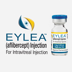
Eylea therapy is a treatment for diabetic retinopathy, retinal vein occlusion and wet-type macular degeneration. It involves repeated painless injections of medication into the eye to prevent blindness by stopping abnormally leaky blood vessels that occur in the eye conditions listed above. Other similar medications that are also used in these conditions include Avastin, Lucentis, Vabysmo, and Beovu.
How effective is Eylea therapy?
Eylea was proven in FDA-approved studies to be effective. In wet-type macular degeneration, monthly or bimonthly injections of Eylea over a one-year period offered a 95% chance of losing less than three lines on a standard eye chart. Eylea was also shown to be effective in the treatment of diabetic retinopathy and retinal vein occlusion to improve vision and prevent severe complications. The results with Eylea are similar to treatment with Lucentis, Avastin, and Beovu. Eylea therapy often starts with injections every 4-6 weeks. Afterwards, the injections may be given less frequently. In some cases the injections may be stopped, but continued monitoring is necessary. There are several medication options apart from Eylea. The best choice of medication may depend on the underlying diagnosis. For example, patient who have glaucoma may have better pressure control while under treatment with Eylea compared with other drugs.
What are the risks of Eylea therapy?
Severe complications are very rare, but risks of Eylea injection include bleeding, inflammation, infection, retinal detachment, cataract, glaucoma, and loss of vision/loss of the eye. The risk of retinal detachment is about 1 in 5,000 injections, but the results of surgical repair are poor. There may be an increased risk of difficultly with future cataract surgery estimated to be about 1% of cases. In these cases the fibers (zonules) that hold the cataract in place may become weaken from Eylea injection. When this occurs, special techniques are required to remove the cataract and place a lens implant. Rarely, two procedures are required to accomplish the task. Studies are ongoing to determine if there may be an increased risk of stroke with Eylea therapy. The possible risk of stroke may be related to the older age of patients with AMD. Further investigation will provide more information. Pregnancy should be avoided while on Eylea therapy.
Intra-vitreal injection
What do I expect after an Eylea injection?
Be careful not to rub the eye after the injection because the eye may remain anesthetized for several hours. You may be given eye drops and instructions on how to use them. Physical activity is not limited after the injection. Tylenol or Ibuprofen may be used if there is discomfort after the injection, but severe pain should be reported to your doctor without delay. It is normal to experience a red area on the white of the eye, which disappears in one to two weeks. If you have any questions or concerns, please call the doctor’s office.
Avastin therapy is a treatment for retinal conditions involving abnormal blood vessel leakage including wet-type age-related macular degeneration, myopic macular degeneration, retinal vein occlusion, diabetic retinopathy, and cystoid macular edema. The treatment involves the painless injection of medication into the eye to stop the leakage and improve vision. The benefits of treatment last one or more months. Repeat injections are common in order to keep the leakage from returning. When the problem has stabilized, the injections may be given less often or discontinued in some conditions. Avastin has not been reviewed by the FDA for use in the eye; therefore, it is used off-label. Safety and effectiveness has been established through extensive experience with the use of Avastin in the eye for a multitude of problems since 2005. There are other similar drugs that have been FDA-approved for use in the eye including Lucentis,Eylea, Vabysmo, and Beovu. These drugs are much more expensive than Avastin. Avastin costs about $50 compared to $2,000 with the FDA-approved drugs. There may be reasons to use one medication over another depending on the diagnosis.
How effective is Avastin therapy?
Avastin has been found to be effective in the treatment of a variety of retinal disorders of blood vessel leakage. It was shown to be as effective as Lucentis in the treatment of wet-type macular degeneration in most patients. Avastin is also effective in the treatment of macular edema, retinal vein occlusion, diabetic retinopathy and other conditions of the eye.
What are the risks of Avastin therapy?
Severe complications are very rare, but risks of Avastin injection include bleeding, infection, inflammation, glaucoma, retinal detachment, cataract, and loss of vision/loss of the eye. The risk of retinal detachment is about 1 in 5,000 injections, but the results of surgical repair are poor. There may be an increased risk of difficultly with future cataract surgery estimated to be about 1%. In these cases the zonules that hold the cataract in place may become weaken from Avastin injection. When this occurs, special techniques are required to remove the cataract and place a lens implant. Rarely, two procedures are required to accomplish the task. Studies are ongoing to determine if there may be an increased risk of stroke with Avastin therapy. Further research is needed. However, pregnancy should be avoided while on Avastin therapy.
There appears to be a greater risk of high eye pressure (glaucoma) in eyes treated with Avastin compared with Lucentis and Eylea. This may be especially important in patients at increased risk of glaucoma due to past high eye pressures or positive family history of glaucoma.
Because Avastin must be measured and placed in a syringe by a compounding pharmacy after manufacture, there may be increased risk of complications with Avastin compared with other similar drugs such as Lucentis, Eylea, Beovu, and Vabysmo. There may be an increased risk of infection due to the introduction of bacteria during repackaging. Some patients experience persistent round floaters due to silicone droplets used to lubricate the syringe from the pharmacy. Over the years, there have been concerns over needle quality (sharpness), which can make injection more uncomfortable.
Intra-vitreal injection
What do I expect after an Avastin injection?
If a patch is placed on the eye, keep it on as directed by the doctor, usually 3-4 hours. You may be given eye drops and instructions on how to use them. Physical activity is not limited after the injection. Tylenol or Ibuprofen may be used if there is discomfort after the injection, but severe pain should be reported to your doctor without delay. It is normal to experience a red area on the white of the eye, which disappears in one to two weeks. If you have any questions or concerns, please call the office.
The Argus II retinal implant is an electronic instrument used to restore limited vision in patients who are blind in both eyes from retinitis pigmentosa. The retina is a thin layer of delicate tissue in the back of your eye, which lines the inside wall like the film in a camera. The retina “takes the picture” of objects and sends the message to the brain. Retinitis pigmentosa is a group of inherited diseases that affects the retina and may cause a profound loss of vision.
How does the Argus II retinal implant work?
The Argus II retinal implant system has several parts. There is a small video camera placed on glasses. The camera records video images and transmits the information to a video processor worn on the belt around the waist. The processor then converts the video information into a digital code that is transmitted to an implant that has been surgically inserted into the eye. The implant includes a set of diodes that are placed inside the eye on the surface of the retina and a coil that is secured to outside of the eye wall underneath the skin where it cannot be seen.
Who is eligible for the Argus II retinal implant?
In February 2013 The FDA granted approval for the use of the Argus II retinal implant only to patients with severe vision loss due to advanced retinitis pigmentosa. Eligible patients must have had good vision early in life and lost all but bare light perception or worse. Patients must also be older than 25 years of age. Researchers hope that with further research the device will be approved in the future for patients with less severe vision loss and for patients with other types of retinal disease. The implant is expected to become available in late 2013.
How much is the vision improved with the Argus II retinal implant?
The improvement in vision is very limited, but helpful in select patients. No clear image is seen. However, eligible patients with the retinal implant are able to see borders between light and dark. This allows them to function better with simple visual tasks such as walking and seeing objects with high contrast. The amount of improvement varies from patient to patient. Because there are risks to surgery, the FDA is appropriately cautious in its approval of the device only for patients with profound loss of vision.
A myopic eye is a near-sighted eye. Without glasses the vision is usually quite good at near, but blurred at distance. Myopia affects 25% of Americans and about 22% of the world population. High myopia (greater than -6.00 diopters) is less common, affecting about 2% of the world population and projected to rise to 10% by the year 2050.
What causes myopia?
Myopia is an inherited condition that usually develops in childhood or early adulthood. The eye, which is round like a ball at birth, becomes oval like an egg. The outer appearance of the eye is not usually changed, but the elongation of the eye changes the focus of the eye from distance to near. There is evidence to suggest that extensive near work (e.g. reading) may worsen myopia.
A myopic eye has elongated somewhat like an egg. Incoming images do not focus on the retina in the back of the eye.
Why is it important to know about myopia?
Although most people with myopia do not develop complications, highly near-sighted people are at increased risk of losing vision from glaucoma, cataract, macular degeneration, and retinal detachment. The higher the degree of near-sightedness (myopia greater than -6.00 diopters), the greater the risk of loss of vision.
Glaucoma is a condition in which the pressure inside the eye damages nerve tissue that helps you see. This pressure usually causes no pain or discomfort and pressure measurements may be normal at times. Over months to years, the pressure slowly takes away the side vision. If undetected and untreated, it may cause total blindness. The best way to diagnose glaucoma is to have regular eye exams each year with pressure measurements. Treatment is effective in preventing vision loss through the use of eye drops. Sometimes, laser or surgery is needed.
Myopic macular degeneration is an uncommon cause of vision loss from severe myopia. The macula is the central part of the retina in the back of the eye. The retina is a thin layer of delicate nerve tissue that lines the inside wall of the eye like the film in a camera. In the eye, light is focused onto the retina, which “takes the picture” and sends the image to the brain. In very near-sighted eyes, the retina becomes stretched as the eye elongates. As a result, the central vision may become blurred or distorted even with proper glasses. Distortion is when straight lines look wavy or crooked. Blood vessels under the macula may bleed causing sudden blurring, blind spot, or distortion. Any of these symptoms should be reported to the eye doctor without delay, as early treatment with medicine injections and/or laser may prevent further loss of vision.
Retinal detachmentis a separation of the retina from the inside wall of the eye. When the retina detaches, it is no longer in proper position inside the eye. Instead, it is like film that has unrolled inside a camera. When this occurs, a camera cannot take a picture. Similarly, when the retina detaches, the eye loses vision. Warning symptoms prior to retinal detachment may include new floaters or brief flashes of light in the side-vision. Later, a dark curtain or shadow slowly starts off to the side and takes away the vision as the retina detaches. Laser or surgery repairs most retinal detachments. It is important to diagnose a retinal detachment early in order to prevent permanent damage to the retina. Report any new floaters, flashes, or loss of side-vision to your eye doctor without delay.
How is myopia treated?
The standard treatment of myopia is to refocus the eye with eye glasses or contact lenses. LASIK and PRK surgery flatten the cornea to focus images onto the retina. Orthokeratology is a controversial method used to flatten the cornea with contacts lenses worn overnight. Lens implants are a more aggressive measure to focus light in highly near-sighted eyes. All of these methods of treatment are simply aimed to focus the vision. They are not designed to correct the underlying problem of elongation of the eye that leads to complications and loss of vision. Diluted atropine eye drops appear to reduce the progression of myopia in an effort to avoid complications of severe elongation of the eye.
Will LASIK surgery help prevent these complications of myopia?
Although LASIK surgery is very effective at flattening the cornea to help eliminate the need for glasses, it does not restore the spherical shape to the eye. Therefore, it is still necessary to be aware of the warning signs of possible complications from myopia.
What are the Do’s and Don’ts?
Using your eyes to read or work at a computer will not weaken them. Avoid intensive rubbing of your eyes. Remember to have your eyes examined once a year. Report the following symptoms to your eye doctor without delay:
Clear vitreous gel fills the eye (click on image to enlarge)
What is Jetrea® injection used for?
Prior to being discontinued, Jetrea therapy was a treatment for retinal conditions involving abnormal pulling of fibers on the retina. Usually due to ageing, fibers which normally lie on the surface of the retina begin to pull on the retina causing a loss of vision. The treatment involved the injection of medication into the eye to cause of release of traction (pulling) on the retina. It may take weeks to months for Jetrea to take effect.
How effective is Jetrea therapy?
In vitreomacular traction syndrome about 40% of cases improve when the traction is limited. In macular hole cases, successful closure of the hole is seen within six months in as many as 60% of eyes with small holes. Please refer to separate literature on these conditions.
Vitreomacular traction relieved by Jetrea (click to enlarge)
What are the risks of Jetrea therapy?
Severe complications are very rare, but risks of Jetrea injection include bleeding, infection, inflammation, glaucoma, dislocation of lens, retinal detachment, cataract, and loss of vision/loss of the eye. A common side effect of treatment is the appearance of new floaters in the vision. Less than one percent of injections are associated with sudden decreased vision for unknown reasons. Fortunately, the vision returns in most cases within a two week period. About 2% of eyes injected with Jetrea experience a yellow tint in the vision which usually clears with time. Currently, it does not appear that Jetrea has any significant systemic adverse effects. However, pregnancy should be avoided while on Jetrea therapy.
What do I expect after a Jetrea injection?
If a patch is placed on the eye, keep it on as directed by the doctor, usually 3-4 hours. You may be given eye drops and instructions on how to use them. Physical activity is not limited after the injection. Tylenol or Ibuprofen may be used if there is discomfort, but severe pain should be reported to your doctor without delay. It is normal to experience a red area on the white of the eye, which disappears in one to two weeks. If you have any questions or concerns, please call the office. If Jetrea is not successful, vitrectomy surgery may be considered.
Note: Since June 30, 2020, Jetrea stopped being manufactured.


